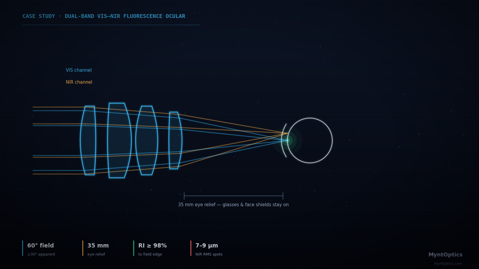
One optical train, two spectral bands: the custom ocular holds visible and NIR channels in register while keeping brightness essentially flat to the field edge.
Application
Near-infrared fluorescence imaging lets clinicians see what white light cannot — perfusion, lymphatics, tissue boundaries — by exciting a fluorophore and imaging its faint NIR emission alongside the normal visible scene. The system this ocular serves presents both channels to the user through a single eyepiece. That puts an unusual burden on a small lens assembly: it must perform across two widely separated spectral bands and present a wide, comfortable, evenly illuminated image, because in fluorescence work a dim image corner is not a cosmetic flaw — it is missing clinical information.
The Challenge
The requirements pulled in four directions at once:
- Dual-band operation: sharp imagery in the visible band (486–656 nm) and the NIR band (830–860 nm) through the same optical train — a chromatic span over which ordinary eyepiece glass choices fall apart.
- Wide apparent field: a full 60° apparent field of view (±30°) for an immersive view of the surgical scene.
- Long eye relief: 35 mm, so the instrument remains usable at a comfortable working posture and with eyewear.
- Uniform brightness: relative illumination had to stay essentially flat to the field edge — the typical cos⁴-driven corner falloff that goes unnoticed in a viewfinder is unacceptable when the signal of interest is photon-starved fluorescence.
Off-the-shelf eyepieces were evaluated first and ruled out with data: none held the combination of eye relief, field and dual-band correction.
What We Analyzed and Delivered
We designed a custom ocular around the client's existing upstream imaging module, treating the supplied modules as fixed black-box elements and engineering the new lens group to complete the system. The design uses anomalous-dispersion glass pairings selected specifically to hold both spectral channels in register, and was optimized with relative illumination as a first-class merit alongside MTF — not as an afterthought to be checked at the end. The full design package included polychromatic MTF for both channels, spot analysis across field, relative illumination curves, and an exit-pupil/interface definition matched to the client's standard mounting.
Achieved Specifications
| Parameter | Achieved |
|---|---|
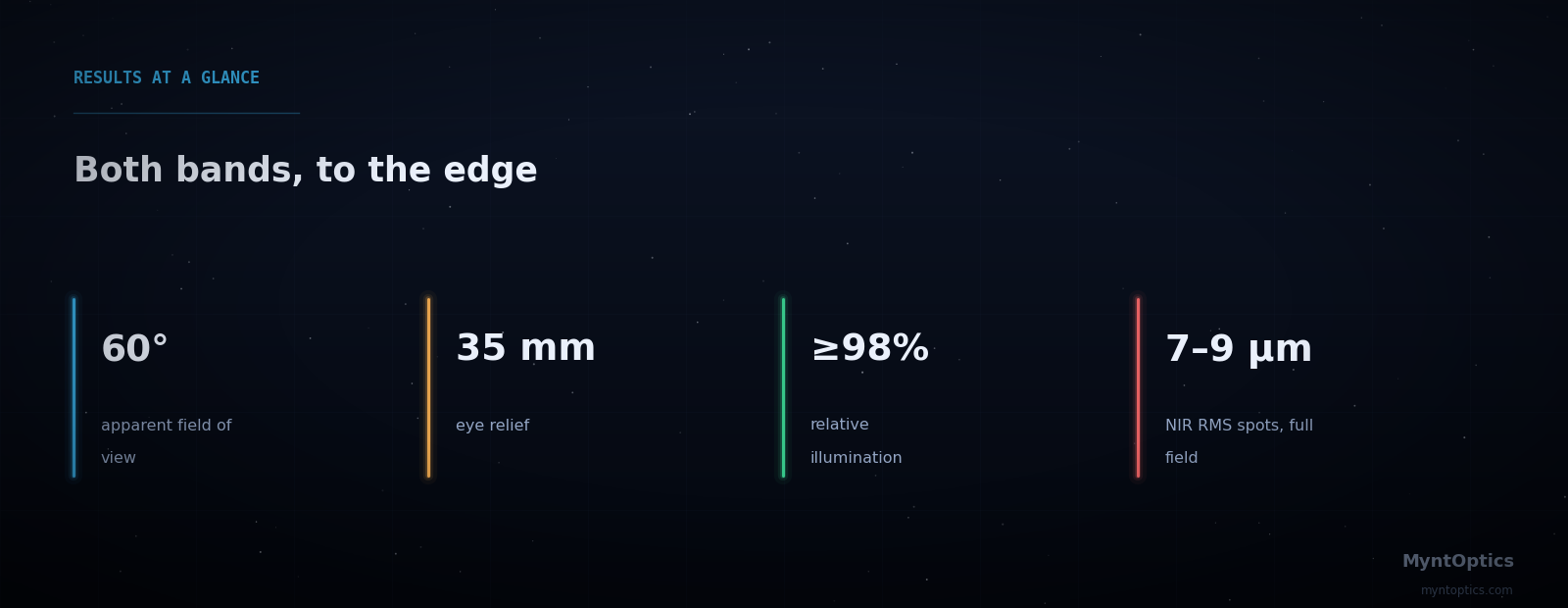
| Apparent field of view | 60° full field (±30°) |
| Eye relief | 35 mm |
| Visible channel | 486–656 nm, near-diffraction-limited core; MTF ≈ 0.5 at 100 cyc/mm at full field |
| NIR channel | 830–860 nm; RMS spot radius 7–9 µm across the full field |
| Relative illumination | ≥98% out to the 30° field edge — visually flat |
| Integration | Designed around existing camera/relay modules; standard mount interface preserved |
Why This Matters
Medical visualization optics are systems problems disguised as component problems. The ocular only succeeds because it was designed with the rest of the instrument — its pupils, its spectral channels, its mechanical interfaces — rather than picked from a catalog and hoped into place. MyntOptics specializes in exactly this kind of constrained, integration-aware design: we work around the hardware you already have, we optimize for the metrics your application actually lives on, and we document the result to medical-device standards of evidence.
Developing an imaging instrument where every photon counts? Talk to MyntOptics about custom visualization optics engineered for your system, not someone else's.
Have a similar engineering challenge? Talk to our optical engineers — a fixed-scope diagnostic turns uncertainty into a costed plan, typically within weeks.