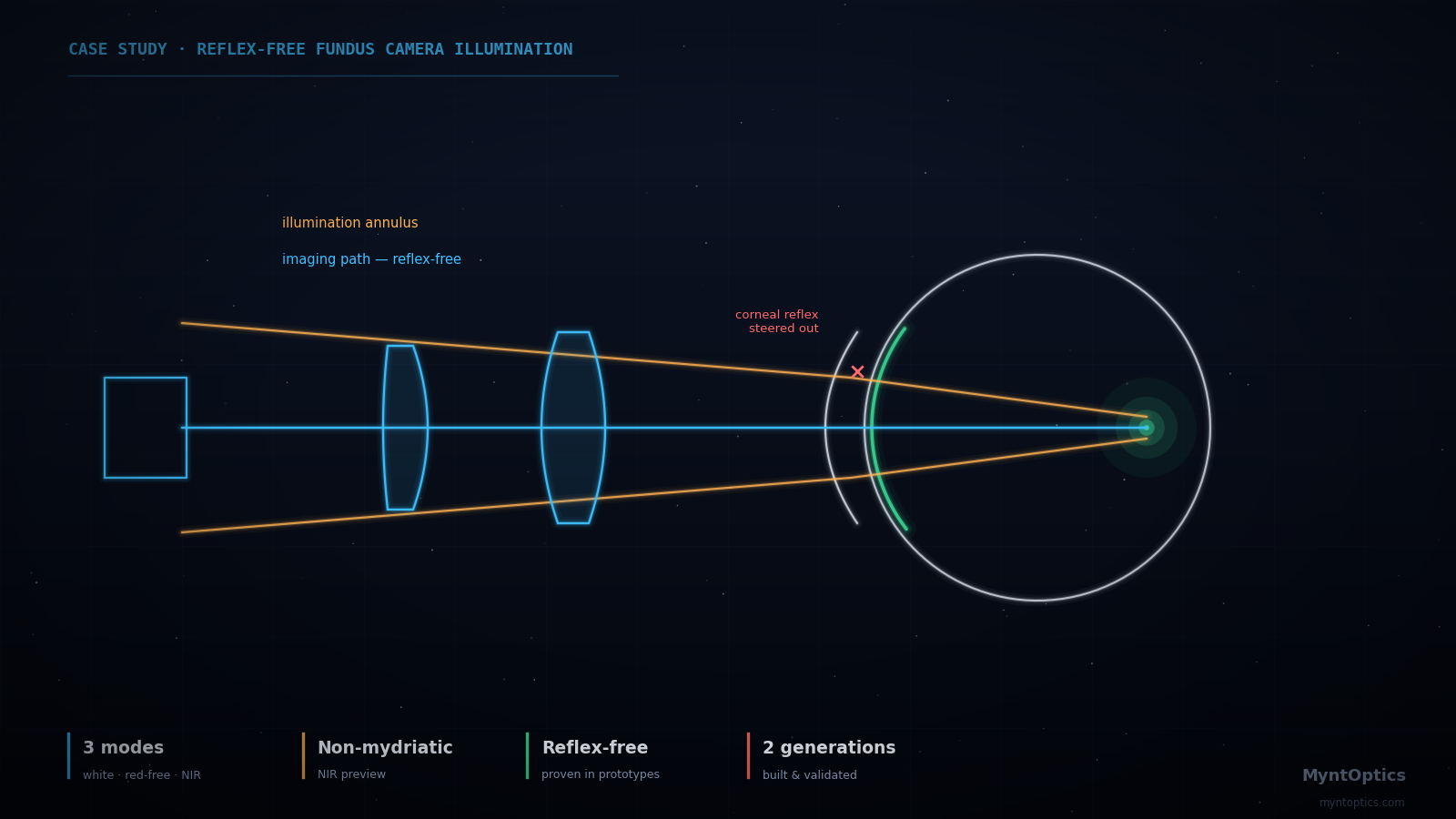
The illumination ring enters the eye around the imaging aperture — so the corneal reflex never reaches the detector.
Application
Retinal screening is one of the highest-leverage diagnostics in medicine — diabetic retinopathy, glaucoma and hypertensive damage all announce themselves at the back of the eye. Bringing that capability out of the specialist clinic requires a fundus camera that is compact, affordable and usable without pupil dilation. This program covered the optical engineering of such a device, from illumination architecture through working prototype.
The Challenge
The fundus camera is a famously self-defeating instrument: the same pupil that admits the illumination must return the image, and the cornea is an excellent convex mirror pointed straight back at the detector. Get the geometry slightly wrong and the image of the retina is buried under corneal glints, crystalline-lens backscatter and internal reflections from the instrument's own optics. In a compact, cost-constrained device, the classical solutions — large annular optics, complex gonio-style illumination — are not available. The illumination and imaging channels had to share a small objective and still stay out of each other's way.
What We Analyzed and Delivered
- Reflex-separated illumination architecture: an illumination geometry that routes source light into the eye while keeping corneal and lens reflexes outside the imaging aperture, developed and verified through systematic stray-light analysis of the full optical train — including the unwanted paths, not just the intended one.
- Multispectral illumination engineering: characterization and integration of white, green and near-infrared LED sources — white for true-color fundus capture, green (red-free) for vessel contrast, NIR for alignment and preview without constricting the patient's pupil, enabling non-mydriatic operation.
- Imaging path design matched to the illumination scheme and packaged for a handheld-class device, with the optical and mechanical trades documented at each iteration.
- Prototype iteration and validation: two prototype generations, with optical performance verified on model eyes and in real capture sessions — culminating in clean, clinically interpretable retinal images with suppressed reflexes.
Achieved Results
| Aspect | Outcome |
|---|---|
| Reflex control | Corneal and lens back-reflections excluded from the imaging channel; verified in prototype imagery |
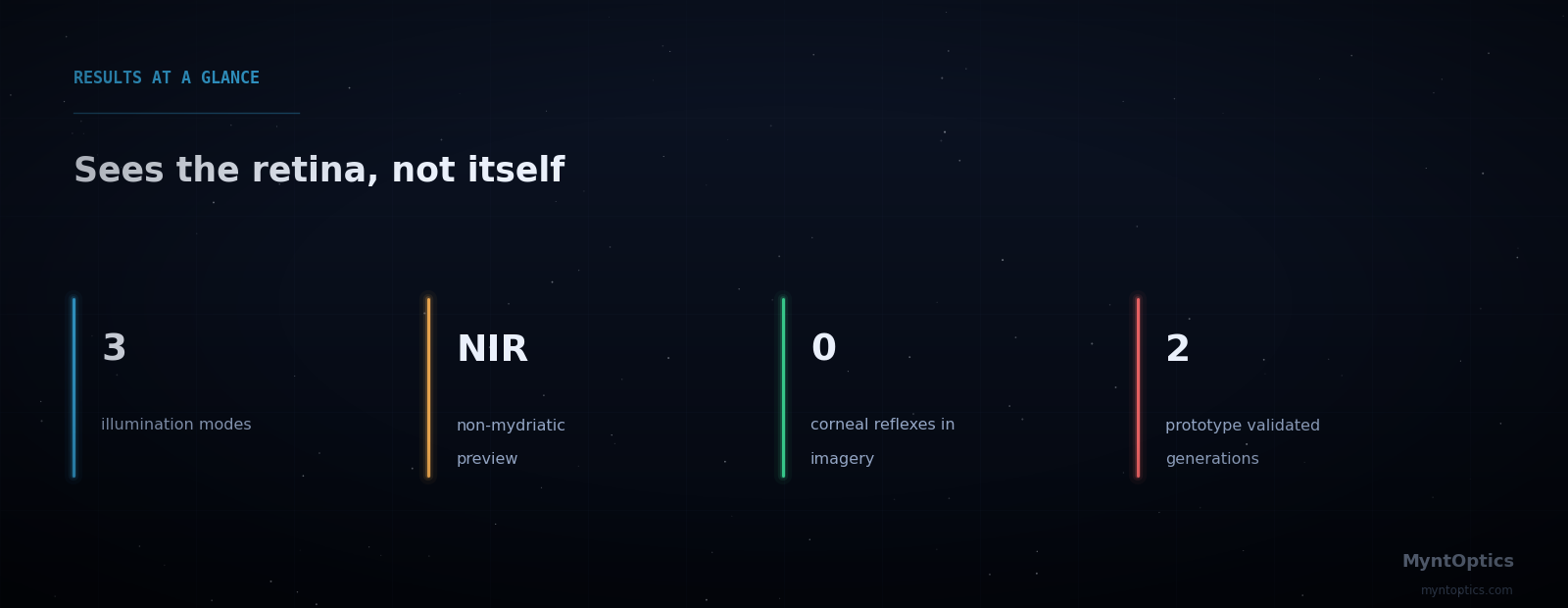
| Illumination modes | White (color fundus), green (red-free vascular contrast), NIR (non-mydriatic alignment) |
| Operation | Non-mydriatic workflow — NIR preview, flash capture |
| Form factor | Compact, low-cost architecture suitable for portable screening use |
| Validation | Working prototypes captured high-quality human retinal images across two design generations |
Why This Matters
Illumination design is the half of imaging-system engineering that rarely appears in brochures and routinely sinks products. A device that images its own light source fails in the field no matter how good its lens MTF is. MyntOptics engineers the illumination and the imaging path as one coupled system — analyzed together, toleranced together, prototyped together — which is why our designs survive contact with real corneas, real patients and real clinics.
Building a diagnostic or imaging device with a difficult illumination problem? Get in touch — this is the part of optics we enjoy most.
Have a similar engineering challenge? Talk to our optical engineers — a fixed-scope diagnostic turns uncertainty into a costed plan, typically within weeks.